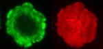
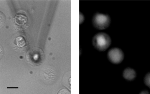
Feeding a Toxoplasma gondii oocyte to a macrophage
Overlay, confocal, of patterned cells
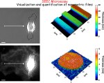
SEEC microscopy : visualization & quantification of nm films
Flow chamber simulations
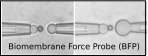
Single molecule measurements of TCR/pMHC interactions on living T cells (Puech et al, PLoS Ones 2011)
Live imaging of actin and membrane on T cells
Jurkat cells as seen using a DualView setup, transfected with FRET based ROZA sensor.
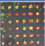
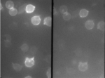
Confocal images of patterned cells
Cover
Passage of a THP-1 leukocyte through a 4 μm wide constriction (Preira et al., Int J Nanotechnol, 2012; Gabriele et al., Lab on a chip 10:1459, 2010; Gabriele et al. Biophys J 96:4308, 2009)
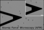
Prototype of diagnostic microfluidic chip to sort cells by size/deformability (Preira et al. Lab Chip 2013)
We code, yes, we code
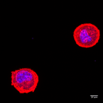
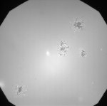
Stimulating calcium probe loaded immune cells (Cazaux et al Ultramicroscopy 2016)
Interference microscopy of T cells on supported lipid bilayers
Cover
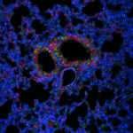
Lung epithelial cells of transgenic mouse expressing the rainbow transgene.
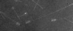
Optical microscopy image in live conditions and without fluorescent staining (taken in Wet-SEEC mode) of the slime deposited in the wake of gliding Myxococcus xanthus bacteria. The thickness of the deposition is less than 5 nm (Ducret et al., PNAS 2012)
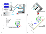
Principe of flow chamber automata and new modalities
Fluorescence imaging on T cells on supported bilayers


You must be logged in to post a comment.