Feeding a Toxoplasma gondii oocyte to a macrophage
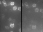
Live imaging of actin and membrane on T cells
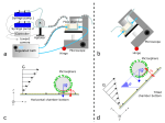
Single molecule measurements of TCR/pMHC interactions on living T cells (Puech et al, PLoS Ones 2011)
Principe of flow chamber automata and new modalities
Jurkat cells as seen using a DualView setup, transfected with FRET based ROZA sensor.
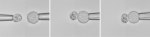
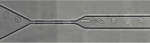
Passage of a THP-1 leukocyte through a 4 μm wide constriction (Preira et al., Int J Nanotechnol, 2012; Gabriele et al., Lab on a chip 10:1459, 2010; Gabriele et al. Biophys J 96:4308, 2009)
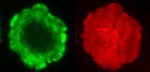
Overlay, confocal, of patterned cells
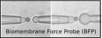
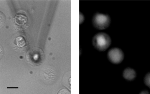
Biomembrane force probe configurations
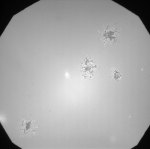
Stimulating calcium probe loaded immune cells (Cazaux et al Ultramicroscopy 2016)
Cover
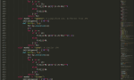
We code, yes, we code
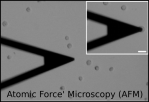
AFM imaging of patterned cells
Interference microscopy of T cells on supported lipid bilayers
Alveole (fluorescent) protein printing
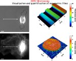
SEEC microscopy : visualization & quantification of nm films
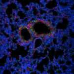
Lung epithelial cells of transgenic mouse expressing the rainbow transgene.
Prototype of diagnostic microfluidic chip to sort cells by size/deformability (Preira et al. Lab Chip 2013)
Cover
Fluorescence imaging on T cells on supported bilayers
Flow chamber simulations
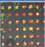
Confocal images of patterned cells
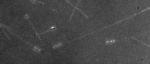
Optical microscopy image in live conditions and without fluorescent staining (taken in Wet-SEEC mode) of the slime deposited in the wake of gliding Myxococcus xanthus bacteria. The thickness of the deposition is less than 5 nm (Ducret et al., PNAS 2012)


You must be logged in to post a comment.