Interactions with surfaces, either artifical or cellular, play a key role in the function of immune cells, and in particular for the T lymphocyte. Using receptor ligand interactions and cytoskeleton deformations, cells are exterting and feeling forces which are essential for the cells to understand the surrounding world, in order to (re)act adequately. This capacity to integrate and evaluate those mechanical signals is called mechanotransduction and its understanding is a major current challenge in cell biology and biophysics. We set to dissect this phenomena at single cell scale by combination of approaches using original engineered substrates coupled to high resolution surface optical microscopy. We have also a strong expertise to exert and record forces at different scales of space and time, using force based biophysical techniques (atomic force microscopy, optical tweezers, micromanipulations, biomembrane force probe, flow chamber). We develop original setups to couple those to recording by fluorescence microscopy the activation state of the lymphocyte using calcium or phosphorylation probes. Using photoactivable molecules, we also directly play on mechanical organization of the cells.
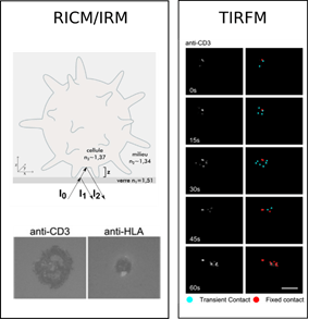 T lymphocyte recognition (P. Bongrand, A. Brodovitch, L. Limozin, A. Pierres, PH. Puech, P. Robert; Collab. C. Boyer (CIML), A Van der Merwe (Oxford)). The detection of foreign material by T lymphocytes is both a key step of immune responses and a model of cell decision triggering. We addressed this model by several parallel approaches based on quantitative advanced biophysical methods. We measured the dynamics of T cell membrane interaction with planar surface with nearly nanometric and subsecond resolution, using reflection interference contrast microscopy (RICM) and evanescent wave illumination (TIRF). We found that T cells could detect the presence of foreign material within less than 10 seconds with highly motile microvilli and pulling motion (J. Immunol. 2013), starting ultimately active spreading, the earliest reporter of bona fide cell activation (J. Immunol. Methods 2011). T cell could quantitatively discriminate between specific antigens of varying potency within 45 seconds after initial contact (Eur. J. Immunol 2015). Quantitative analysis of TIRF images revealed that T cell plasma membranes displayed transverse undulations of several tens of nanometer amplitude at one hertz frequency (Cell Mol Bioengineering, 2015), an efficient way of mechanosensing the nearby surface through the TCR. The importance and significance of addressed problems was discussed in two review papers (Frontiers Immunol. 2012; Annual Rev. Immunol. 2015).
 Biosensors in T cell activation/migration (A Lellouch, continued by M Biarnes-Pelicot) (Biophys J 2014, Biotech. J 2014, BBRC 2015). We determined that ZAP-70 is activated upon LFA-1 engagement during T cell migration. Furthermore, we demonstrated that the kinase is involved in regulating the recruitment of talin to high affinity LFA-1 and that this process requires binding of the ZAP-70 SH2 domains to a yet to be identified partner. Aside of those reporters, we developped photoactivable proteins, such as PA-Rac, in order to use the power of optogenetics to perturb cell cytoskeleton and function (Ultramiscroscopy 2016).
 Surface control of T lymphocyte adhesion (L. Limozin, P. Dillard, AM Varma (NIH)). We interrogate T lymphocyte physiology by designing carefully controlled artificial surfaces to stimulate cell activation and spreading. Our original artificial surfaces exhibit fixed or mobile ligands, and arrays of submicrometric adhesive patches surrounded with repulsive zones (Nanoletters 2013, 2015). These surfaces are compatible with high resolution surface microscopy like TIRFM and RICM. With these tools, we have demonstrated that (1) ligand mobility controls lymphocyte spreading and we proposed a physical mechanism based on friction to explain these results (Biophys. J. 2014), (2) cell adhesion area is determined by global ligand density, but local cell-substrate interface and receptor clustering is tuned by underlying adhesive patch geometry (Integr. Biol. 2016).
Coupling AFM force mode and fluorescence microscopy for mechanotransduction studies (PH Puech, A. Sadoun, collab. Y. Hamon, CIML). We designed a simple method to mechanically stimulate immune cells while recording calcium pulses, and observe T cell mechanical response upon photoactivation of a small G protein (Ultramicroscopy 2016).


You must be logged in to post a comment.