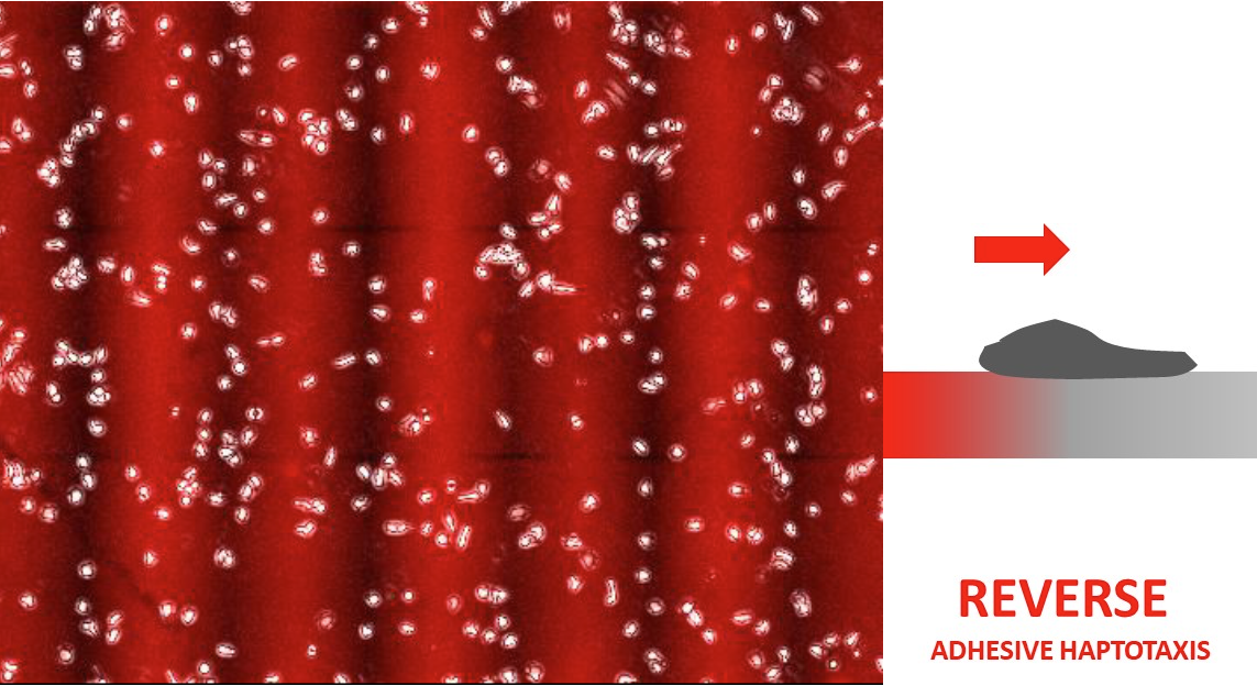
Cell guidance by anchored molecules, or haptotaxis, is crucial in development, immunology and cancer. Adhesive haptotaxis, or guidance by adhesion molecules, is well established for mesenchymal cells such as fibroblasts, whereas its existence remains unreported for amoeboid cells that require less or no adhesion in order to migrate. We show that, in vitro, amoeboid human T lymphocytes develop adhesive haptotaxis mediated by densities of integrin ligands expressed by high endothelial venules. Moreover, lymphocytes orient towards increasing adhesion with VLA4 integrins (also known as integrin α4β1), like all mesenchymal cells, but towards decreasing adhesion with LFA-1 integrins (also known as integrin αLβ4), which has not previously been observed. This counterintuitive ‘reverse haptotaxis’ cannot be explained by existing mechanisms of mesenchymal haptotaxis involving either competitive anchoring of cell edges under tension or differential integrin-activated growth of lamellipodia, because they both favor orientation towards increasing adhesion. The mechanisms and functions of amoeboid adhesive haptotaxis remain unclear; however, multidirectional integrin-mediated haptotaxis might operate around transmigration ports on endothelia, stromal cells in lymph nodes, and inflamed tissue where integrin ligands are spatially modulated.
Researchers from LAI and CINAM have joined their efforts to study how T-lymphocytes sense the mechanics of their underlying substrate in vitro, an important but poorly understood question. Using a wide range of stiffnesses for substrates covered with antibodies against T-Cell receptors, they observed that T-cell spread more on increasingly stiff substrates, until a maximal stiffness, where they start to spread less. This is reconciling apparent contradictory results from the literature. It was also observed that involvement of integrins in this process reestablish a monotonous response, as observed for non-immune cells. The observations are rationalized in terms of a simple quantitative clutch model which will be further tested.
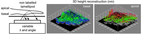
Long-time expertise of LAI (in collaboration with CINAM) in Reflection interference contrast microscopy have recently permitted a new advance to perform nanometer scale mapping of the cell lamellipod in vitro. Combining multicolor RICM with in silico reconstruction and using optical modeling, the topography of the upper and lower membrane of a lamellipod was reconstructed, as well as the refractive index mapped. These measurements were successfully compared with independent determinations using Atomic Force Microscopy or Quantitative Phase Imaging which both provide only a partial information.
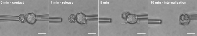
Toxoplasma gondii is a common parasite of humans and animals, which is transmitted via oocysts in cat faeces or tissue cysts in contaminated meat. Given the resistance of the oocyst wall to digestive enzymes and the ability of oocysts to cause parenteral infections, the present study investigated the possible contribution of macrophages in supporting sporozoite excystation following oocyst internalisation. By using single cell micromanipulations, real-time and time-point imaging techniques, we demonstrated that RAW macrophages could interact rapidly with oocysts and engulfed them by remodelling of their actin cytoskeleton. Internalised oocysts were associated to macrophage acidic compartments and showed evidences of wall disruption. Sporozoites were observed in macrophages containing oocyst remnants or in new macrophages, giving rise to dividing tachyzoites. We highligh an unexpected role for the macrophage in facilitating the excystation and differentiation of T. gondii sporozoites following oocyst internalisation.
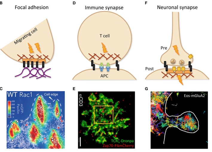
The plasma membrane delimits the cell, which is the basic unit of living organisms, and is also a privileged site for cell communication with the environment. Cell adhesion can occur through cell-cell and cell-matrix contacts. Adhesion proteins such as integrins and cadherins also constitute receptors for inside-out and outside-in signaling within proteolipidic platforms. Adhesion molecule targeting and stabilization relies on specific features such as preferential segregation by the sub-membrane cytoskeleton meshwork and within membrane proteolipidic microdomains. This review presents an overview of the recent insights brought by the latest developments in microscopy, to unravel the molecular remodeling occurring at cell contacts. The dynamic aspect of cell adhesion was recently highlighted by super-resolution videomicroscopy, also named videonanoscopy. By circumventing the diffraction limit of light, nanoscopy has allowed the monitoring of molecular localization and behavior at the single-molecule level, on fixed and living cells. Accessing molecular-resolution details such as quantitatively monitoring components entering and leaving cell contacts by lateral diffusion and reversible association has revealed an unexpected plasticity. Adhesion structures can be highly specialized, such as focal adhesion in motile cells, as well as immune and neuronal synapses. Spatiotemporal reorganization of adhesion molecules, receptors, and adaptors directly relates to structure/function modulation. Assembly of these supramolecular complexes is continuously balanced by dynamic events, remodeling adhesions on various timescales, notably by molecular conformation switches, lateral diffusion within the membrane and endo/exocytosis. Pathological alterations in cell adhesion are involved in cancer evolution, through cancer stem cell interaction with stromal niches, growth, extravasation, and metastasis.
As depicted by cartoons (B,D,F) and illustrated by experimental data (C,E,G), specialized cell contacts can be implicated in structures such as focal adhesion (B,C), immune (between T cell and APC; D,E), and neuronal (between pre- and post-synaptic neurons) synapses (F,G), dealing with specific dynamics in relation with their function. (C) Trajectories of wild-type (WT) Rac1, tagged with Halo-tetra-methyl-rhodamin, obtained by single-particle tracking (white lines) and superimposed on mGFP-Paxillin staining (false colors identifying FAs) reveal transient (red dots) or stable (white dots) immobilization within FAs. Reprinted from Shibata et al. (2013). (E) PALM imaging was performed with two molecules of the TCR complex, tagged with photoactivatable fluorescent proteins, TCRζ–Dronpa and ZAP-70-PAmCherry, in an E6.1 Jurkat cell on αCD3-coated coverslip. Nanoscopy of the immune synapse reveals TCR micro- and nano-clusters (green) with ZAP-70 sub-clusters (red) associated to activated TCR. Bar: 2 μm. Reprinted from Neve-Oz et al. (2015). (G) Trajectories of the tagged AMPA receptor Eos-GluA2 measured by sptPALM report transient organization in nanodomains within an excitatory dendritic spine (delimited by the white line) of a rat hippocampal neuron.


You must be logged in to post a comment.