Long-time expertise of LAI (in collaboration with CINAM) in Reflection interference contrast microscopy have recently permitted a new advance to perform nanometer scale mapping of the cell lamellipod in vitro. Combining multicolor RICM with in silico reconstruction and using optical modeling, the topography of the upper and lower membrane of a lamellipod was reconstructed, as well as the refractive index mapped. These measurements were successfully compared with independent determinations using Atomic Force Microscopy or Quantitative Phase Imaging which both provide only a partial information.
https://doi.org/10.1021/acs.nanolett.8b03134
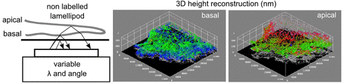
3D representations of basal membrane (left: seen from under) and of apical membrane (right : seen from top).

You must be logged in to post a comment.