European Thymus Meeting Participation

We are excited to share Arnauld Sergé‘s participation in European Thymus in Porto, 27-31 May 2024

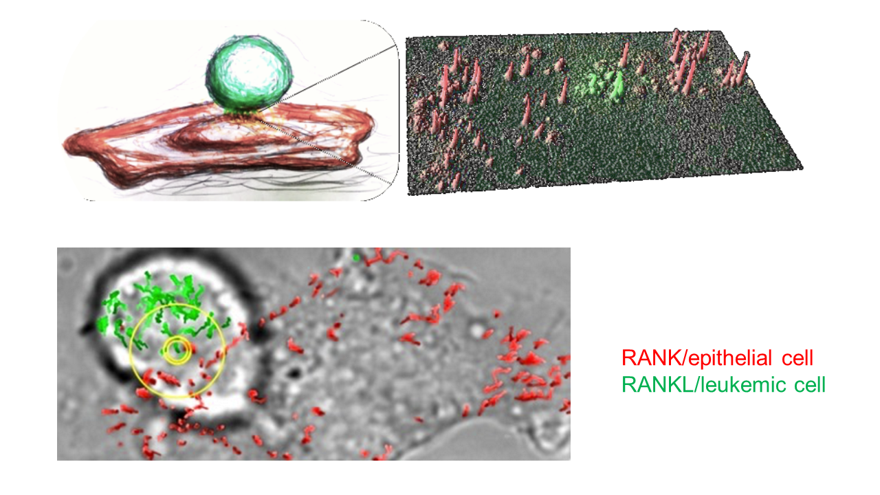
Arnauld presented a poster titled “Dynamics and Blockade of RANK/RANKL Interactions Between Leukemic and Thymic Epithelial Cells,” with contributions from Loriane Maillot, Martine Biarnes Pelicot, and Magali Irla from CIML.

T-cell acute lymphoblastic leukemias (T-ALL) are aggressive hematological cancers arising in the thymus from the malignant transformation of thymocytes. RANK and its ligand RANKL play a key role in thymic crosstalk. RANKL promotes thymic epithelial cell differentiation, sustaining the generation of a self-tolerant T-cell repertoire. The RANK/RANKL axis is a therapeutic target in oncology. However, its role in T-ALL remains unclear. RANKL is overexpressed during leukemogenesis. (i) we evaluate the dynamics of RANK/RANKL interactions, measured by single-molecule tracking. We evaluate the effects of an anti-RANKL antibody, a decoy receptor and the soluble form of RANKL. (ii) we assessthe therapeutic potential of an anti-RANKL antibody in T-ALL dissemination. Our aim is to shed light on the role of the RANK/RANKL axis in the emergence and dissemination of T-ALL.

CENTURI – Turing Centre for Living Systems Scientific Day !

We are pleased to share that Ahmad Awada delivered an insightful presentation titled “Effects of Adhesion and Confinement on Leukocyte Migration” at the CENTURI – Turing Centre for Living Systems scientific day: Success Stories and New Horizons, held on May 30th at Jardins du Pharo.

The event was a remarkable gathering, bolstering the community spirit through networking and the sharing of ideas and future outlooks. It provided an excellent platform for researchers to connect, collaborate, and envision new horizons in scientific research.

LAI celebrated its newest Dr

On the 4th of April 2024, Rémy Torro, Ph.D. successfully defended his work titled “High throughput analysis of dynamic single cell interactions imaged by multimodal optical microscopy” in front of a jury composed of experts in the field, Alice Nicolas, David Rousseau, Daniel Sage, Loïc Dupré under the supervision of Laurent Limozin and Kheya sengupta. His work focused on develloping a software called Celldetective to describe complex cell interactions from multimodal microscopy images, leveraging recent AI techniques and applying it to test cell engagers for immunotherapy in cell-surface and cell-cell interaction assays.

Celldetective is now available and free to use on GitHub: https://lnkd.in/dGpj_UUx

Best of luck, Remy!

remy'defense

In association with CENTURI – Turing Centre for Living Systems

LAI celebrates Marc Karnat’s thesis (Visiting PhD Student from CPT)

Marc did his Ph.D. under the supervision of Jean-Francois Rupprecht at CPT (kindly hosted at LAI) and funded by CENTURI. On monday, 18th December, he successfully defended his thesis which is entitled “Inferring the rheology of cellular materials through dedicated deep learning tools” in front of an expert jury consisting of Romain Levayer, Hervé Turlier, Hélène Delanoë-Ayari, Bianca Habermann, Thierry Artières and Sham Tlili.

Luckily, he’ll stay with us for a few months in 2024 to finish his project and we wish him the best of luck in his future. Again, Congratulations Marc for your incredible work !!

Congratulations Martine Biarnes Pelicot (our amazing engineer) for your contribution to the book “Etonnante physique” published by CNRS !!

We are delighted to inform you that our engineer, Martine Biarnes Pelicot, has contributed to the book “Etonnante physique” published by CNRS. In her article, Martine explains how genetic engineering enables her to create tailor-made tools for studying the biophysics of immune cells.

This book brings together 70 contributions from physicists who have recently been awarded a CNRS medal. The book offers a captivating exploration of the many areas covered by this surprising discipline. It is designed to be accessible to all those who are curious about science.

We invite you to discover this fascinating book by following the link below: https://anneedelaphysique.cnrs.fr/ressource/etonnante-physique/”

A new article on Naive T lymphocytes chemotax long distance to CCL21 but not to a source of bioactive S1P in Iscience

Naive T lymphocytes traffic through the organism in search for antigen, alternating between blood and secondary lymphoid organs. Lymphocyte homing to lymph nodes relies on CCL21 chemokine sensing by CCR7 receptors, while exit into efferent lymphatics relies on sphingolipid S1P sensing by S1PR1 receptors. While both molecules are claimed chemotactic, a quantitative analysis of naive T lymphocyte migration along defined gradients is missing. Here, we used a reductionist approach to study the real-time single-cell response of naive T lymphocytes to CCL21 and serum rich in bioactive S1P. Using microfluidic and micropatterning ad hoc tools, we show that CCL21 triggers stable polarization and long-range chemotaxis of cells, whereas S1P-rich serum triggers a transient polarization only and no significant displacement, potentially representing a brief transmigration step through exit portals. Our in vitro data thus suggest that naive T lymphocyte chemotax long distances to CCL21 but not toward a source of bioactive S1P.

https://doi.org/10.1016/j.isci.2023.107695

Non-exhaustive model of naive T lymphocyte traffic after accessing lymph nodes. Cells are gently attracted by long-range CCL19 and CCL21 gradients toward the central parenchyma, where they encounter higher and homogeneous concentrations of CCL21 that allow their random walk throughout the T cell zone. Moreover, S1P-rich serum triggers a transient polarization only and no significant displacement, potentially representing a brief transmigration step through exit portals.

A new article on living cells guided against a flow by a roly-poly-like mechanism in PNAS

Cellular guidance by chemical or physical signals is essential for many life processes and usually relies on sophisticated biological processes that are still partially elucidated. Microfluidic experiments and mechanical modeling has revealed that the choice for cells to orient themselves against or in the direction of a flow can result from a simple physical bias. They have worked with  keratocytes, cells that form the scale of fishes, and whose morphology is characterized by broad  flat “front” and a compact protruding front  “back”. A simplified model of a cell with a hemispherical back and a flat rectangular front allows to quantitatively calculate the forces that the flow exerts on each edge. The resulting force stabilizes the cells with a large rear edge against the flow, like a roly-poly that stays upright because of its heavy bottom edge. The researchers’ model successfully predicted the experimentally observed orientation for each cell without adjusting parameter. It is an elegant example where a characterized biological behavior does not result from specialized molecular sensors and a complex cascade of internal biosignals to reorient the cell, but from a simple passive physical bias.

https://www.pnas.org/doi/10.1073/pnas.2210379119

A– Keratocytes descending a flow, the white arrow indicates the direction of the stream. B– Cell morphology seen in 3D by confocal microscopy, with a bulbous back edge and a flat, thin front edge. C– Cell modeling with a hemisphere at the back (red) and a flat rectangle at the front (brown) D– The torques resulting from flow on cell front (red arrow) and cell front (brown arrow) stabilize upstream orientation of cell with larger rear edge, like the torque resulting from gravity stabilizes the standing position of roly-poly toys with large bottom edge. 

This paper was commented on in CNRS (link)

A new article on leukemic stem cell adhesion to bone marrow stromal cells by single-molecule tracking nanoscopy in Journal of Cell Science

The interactions between haematopoietic and stromal cells are profoundly altered by leukaemias, contributing to the phenomena of resistance to myeloablative treatments. In this study, we followed the dynamics of JAM adhesion molecules at the membrane between leukaemic and stromal cells by videonanoscopy in order to study the establishment and evolution of these cellular junctions. The trajectories of JAMs were analysed with near-nanometer precision using a dedicated MTT (Multi-Target Tracing, Sergé et al. Nature Methods 2008) algorithm extended to 2 colours, which allows to reveal the signature of interaction and stabilization events at cell contacts. We have thus characterised the involvement of JAMs in the interaction mechanisms of tumour cells as well as the maintenance of stem cells in bone marrow niches through enhanced interaction. From a therapeutic perspective, we destabilised leukaemic stem cells using blocking antibodies opening opportunities for disrupting LSC resistance mechanisms.

https://doi.org/10.1242/jcs.258736

(B) Maximum projection of a 500-frame videonanoscopy acquisition, showing JAM-B and JAM-C positions over time (left). Maps of JAM-B and JAM-C trajectories represented by gradients of green and magenta, respectively, according to time, and superimposed on the transmission image of the cells (right). Inserts show magnifications of the framed areas. Spatiotemporal colocalizations are denoted by white circles with a size proportional to duration. Several concentric circles correspond to successive colocalizations at a nearby locations but with different durations. (C) Images from the same videonanoscopy acquisition corresponding to the area framed in B, with colocalization events or not (white circle or green/magenta arrowheads, respectively).

New article on early leukocyte activation triggered by viscoelastic changes in Biophysical journal

This article presents the evidence that immune cells are regulating very rapidely, even before classical signalling times as recorded by calcium fluxes, their mechanical properties when encountering an activating substrate, being either artificial (bead) or physiological (APC). For this, we developed a micropipette-based rheometer to track cell viscous and elastic properties. We have shown that leukocytes become up to 10 times stiffer and more viscous during their activation. Elastic and viscous properties evolve in parallel, preserving a ratio characteristic of the leukocyte subtype. These mechanical measurements set up a complete picture of the mechanics of leukocyte activation and provide a signature of cell function

https://doi.org/10.1016/

Activation of three types of leukocytes studied with the micropipette rheometer. Left (a): T cell, middle (b): B cell, right (c): PLB cells. All bars represent 5 μm.

A new article on lymphocyte swimming in Biophysical Journal 

SIGNIFICANCE Leukocytes have a ubiquitous capacity to migrate on or in solid matrices and with or without adhesion, which is instrumental to fight infections. The precise mechanisms sustaining migration remain, however, arguable. It is for instance widely accepted that leukocytes cannot crawl on two-dimensional substrates without adhesion. In contrast, we showed that human lymphocytes swim on nonadherent two-dimensional substrates and in suspension. Furthermore, our experiments and modeling suggest that propulsion hardly rely on cell body deformations and predominantly on molecular paddling by transmembrane proteins protruding outside the cell. For physics, this study reveals a new type of microswimmer, and for biology, it suggests that leukocyte’s ubiquitous crawling may have evolved from an early machinery of swimming shared by various eukaryotic cells.

Biophysical Journal 119, 1157–1177, September 15, 2020 1157


This paper was commented on in Science (link), CellPress (link), Eurekalert (link) and Science&Vie (link) and CNRS (link).