The interactions between haematopoietic and stromal cells are profoundly altered by leukaemias, contributing to the phenomena of resistance to myeloablative treatments. In this study, we followed the dynamics of JAM adhesion molecules at the membrane between leukaemic and stromal cells by videonanoscopy in order to study the establishment and evolution of these cellular junctions. The trajectories of JAMs were analysed with near-nanometer precision using a dedicated MTT (Multi-Target Tracing, Sergé et al. Nature Methods 2008) algorithm extended to 2 colours, which allows to reveal the signature of interaction and stabilization events at cell contacts. We have thus characterised the involvement of JAMs in the interaction mechanisms of tumour cells as well as the maintenance of stem cells in bone marrow niches through enhanced interaction. From a therapeutic perspective, we destabilised leukaemic stem cells using blocking antibodies opening opportunities for disrupting LSC resistance mechanisms.
https://doi.org/10.1242/jcs.258736
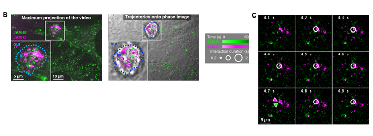
(B) Maximum projection of a 500-frame videonanoscopy acquisition, showing JAM-B and JAM-C positions over time (left). Maps of JAM-B and JAM-C trajectories represented by gradients of green and magenta, respectively, according to time, and superimposed on the transmission image of the cells (right). Inserts show magnifications of the framed areas. Spatiotemporal colocalizations are denoted by white circles with a size proportional to duration. Several concentric circles correspond to successive colocalizations at a nearby locations but with different durations. (C) Images from the same videonanoscopy acquisition corresponding to the area framed in B, with colocalization events or not (white circle or green/magenta arrowheads, respectively).

You must be logged in to post a comment.