Guillou L, Babataheri A, Puech PH, Barakat AI, Husson J (2016) Dynamic monitoring of cell mechanical properties using profile microindentation. Sci. Reports 6:21529.
Article
A new article on spreading on nanostructured surfaces
Dillard P, Pi F, Lellouch AC, Limozin L, Sengupta K (2016) Nano-clustering of ligands on surrogate antigen presenting cells modulates T cell membrane adhesion and organization. Integrative Biology 8:287.
A new article on SDRA / microfluidics
Preira P, Forel J-M, Robert P, Nègre, Biarnes M, Xeridat F, Bongrand P , Papazian L, Théodoly O (2016) The leukocyte stiffening property of plasma in acute respiratory distress syndrome (ARDS) revealed by microfluidic single-cell study: role of cytokines and protection with antibodies. Critical Care 20 : 8.
A new article on medical applications
Bertin D, Mouhajir Y, Bongrand P, Bardin N (2016) ICARE improves antinuclear antibody detection by overcoming the barriers preventing accreditation. Clinica Chimica Acta 454 : 57-61.
A new article on molecular architecture of cell Adhesion by videonanoscopy in Frontiers
The plasma membrane delimits the cell, which is the basic unit of living organisms, and is also a privileged site for cell communication with the environment. Cell adhesion can occur through cell-cell and cell-matrix contacts. Adhesion proteins such as integrins and cadherins also constitute receptors for inside-out and outside-in signaling within proteolipidic platforms. Adhesion molecule targeting and stabilization relies on specific features such as preferential segregation by the sub-membrane cytoskeleton meshwork and within membrane proteolipidic microdomains. This review presents an overview of the recent insights brought by the latest developments in microscopy, to unravel the molecular remodeling occurring at cell contacts. The dynamic aspect of cell adhesion was recently highlighted by super-resolution videomicroscopy, also named videonanoscopy. By circumventing the diffraction limit of light, nanoscopy has allowed the monitoring of molecular localization and behavior at the single-molecule level, on fixed and living cells. Accessing molecular-resolution details such as quantitatively monitoring components entering and leaving cell contacts by lateral diffusion and reversible association has revealed an unexpected plasticity. Adhesion structures can be highly specialized, such as focal adhesion in motile cells, as well as immune and neuronal synapses. Spatiotemporal reorganization of adhesion molecules, receptors, and adaptors directly relates to structure/function modulation. Assembly of these supramolecular complexes is continuously balanced by dynamic events, remodeling adhesions on various timescales, notably by molecular conformation switches, lateral diffusion within the membrane and endo/exocytosis. Pathological alterations in cell adhesion are involved in cancer evolution, through cancer stem cell interaction with stromal niches, growth, extravasation, and metastasis.
https://doi.org/10.3389/fcell.2016.00036
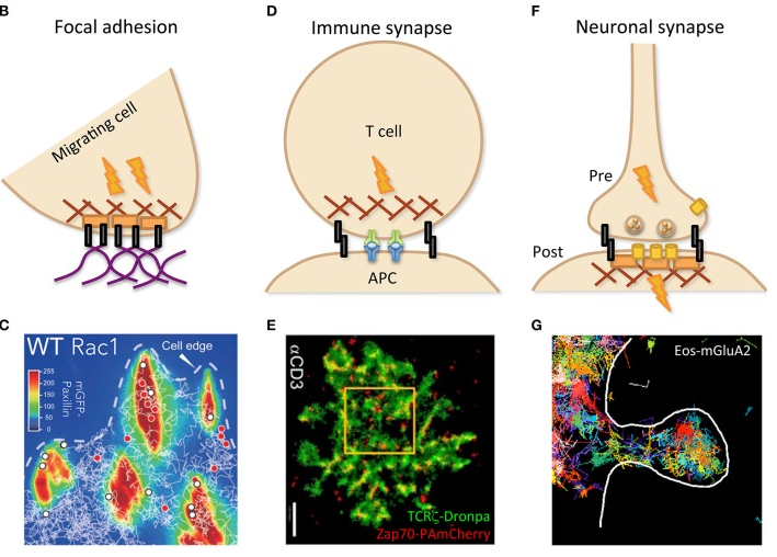
As depicted by cartoons (B,D,F) and illustrated by experimental data (C,E,G), specialized cell contacts can be implicated in structures such as focal adhesion (B,C), immune (between T cell and APC; D,E), and neuronal (between pre- and post-synaptic neurons) synapses (F,G), dealing with specific dynamics in relation with their function. (C) Trajectories of wild-type (WT) Rac1, tagged with Halo-tetra-methyl-rhodamin, obtained by single-particle tracking (white lines) and superimposed on mGFP-Paxillin staining (false colors identifying FAs) reveal transient (red dots) or stable (white dots) immobilization within FAs. Reprinted from Shibata et al. (2013). (E) PALM imaging was performed with two molecules of the TCR complex, tagged with photoactivatable fluorescent proteins, TCRζ–Dronpa and ZAP-70-PAmCherry, in an E6.1 Jurkat cell on αCD3-coated coverslip. Nanoscopy of the immune synapse reveals TCR micro- and nano-clusters (green) with ZAP-70 sub-clusters (red) associated to activated TCR. Bar: 2 μm. Reprinted from Neve-Oz et al. (2015). (G) Trajectories of the tagged AMPA receptor Eos-GluA2 measured by sptPALM report transient organization in nanodomains within an excitatory dendritic spine (delimited by the white line) of a rat hippocampal neuron.
A new article on AFM / fluorescence
Cazaux S, Sadoun A, Pelicot-Biarnes M, Martinez M, Obeid S, Bongrand P, Limozin L, Puech P-H (2016) Synchronizing atomic force microscopy force mode and fluorescence microscopy in real time for immune cell stimulation and activation studies. Ultramicroscopy 160: 168-181.
Microfluidics, cell rheology and medical transfer
The long term effort to develop microfluidic tools to probe the rheological properties of circulating leukocytes has allowed getting new insight in the triggering mechanisms of the acute respiratory distress syndrome. This work, performed in a collaboration with Dr Forel and Pr Papazian from AP-HM has been published in Critical Care: “The leukocyte-stiffening property of plasma in early acute respiratory distress syndrome (ARDS) revealed by a microfluidic single-cell study: the role of cytokines and protection with antibodies”
Critical Care (2016) 20:8 DOI 10.1186/s13054-015-1157-5

Cell self-steer like single-handed circumnavigators

The mechanism of lymphocytes guiding against a flow was shown to use the tail of lymphocyte like a wind vane in a breeze. This wind vane self-steering system is reminiscent ofa system used by sailors to orient their ship with the wind.
This interdisciplinary work between physics, biology and yachting was published in Nature communication: “Lymphocytes can self-steer passively with wind vane uropods” By:Valignat, MP, Negre, P, Cadra, S, Lellouch, AC, Gallet, F, Henon, S, Theodoly, O;NATURE COMMUNICATIONS, 5, 5213, DOI: 10.1038/ncomms6213, OCT 2014.
See also on the CNRS web site (INP ‘Actualités)

You must be logged in to post a comment.