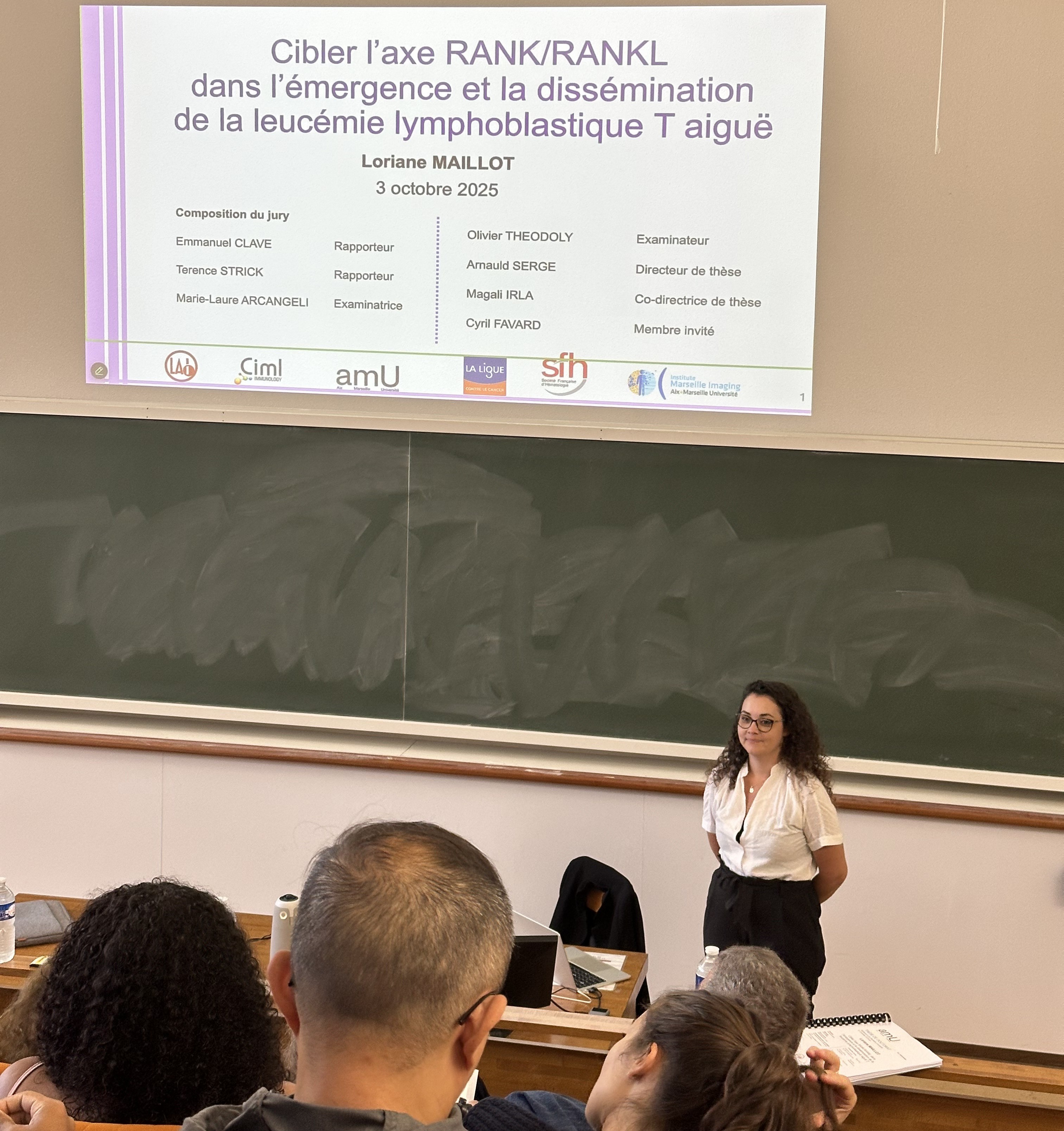
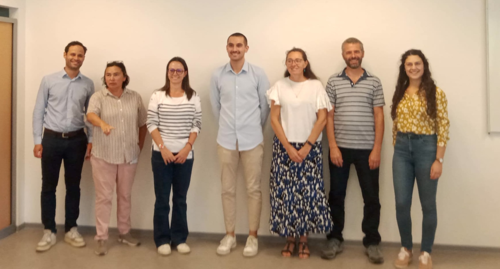
We are pleased to share that Jana El Husseiny presented her work “Unraveling Morphological and Biomechanical Responses to Stressors in Coccidian Oocysts”, and Gaurav Verma gave a flash talk titled “Molecular Geometry Controls Immune Response” at the CENTURI – Turing Centre for Living Systems Scientific Day, held on September 17th at Jardins du Pharo.
The event brought together current members and alumni, fostering networking, idea exchange, and discussions on future perspectives. The day concluded with a convivial cocktail evening at Villa Gaby.
On the 16th of september 2025, Chandrasekar Subramani Narayana successfully defended his work titled “Deep learning-based counting and binding kinetics of bacteriophages imaged by fluorescence microscopy“, under the supervision of Laurent Limozin and Stéphane Ayache. This achievement was presented to a distinguished panel of experts in the field, including Laurent Vonna, Rémi Eyraud, Guillaume Gay and Cécile Capponi,
LAI wishes you all the best for the rest of your career, full of success, and thanks you for your hard work and the positive energy you shared within the laboratory.
We present a detailed protocol for conducting high-content microscopy assays of immune cells interacting with antigen surfaces and antigen-presenting cells. The principle of the method is illustrated by redirecting NK cells on HER2 antigens using original bispecific antibodies. We provide a complete analysis pipeline using our open-source, home-made software, Celldetective.
We are very proud of Dr. Zacharie Nakache, who has defended his PhD in Pharmacy, performed under the co-supervision of Prof. Arnauld Sergé at LAI and Dr. Magali Irla at CIM
On the 23th of June 2025, Luc David Broglio successfully defended his work titled “Outils diagnostics pour l’immunologie basés sur la micro-impression d’anticorps“, under the supervision of Olivier Theodoly and Philippe Robert. This achievement was presented to a distinguished panel of experts in the field, including Charlotte Riviere, Sylvain Gabriele, Régis Costello, Sophie Asnacios and Vincent Studer.
We thank Luc for his dedicated work developing microchips printed with multiple functional proteins to measure the kinetics of leukocyte activation at the single-cell level. The entire lab warmly congratulates Luc for this important milestone and wishes him continued success in his future scientific endeavors!
We were delighted to host Jay Groves from the University of California, Berkeley, during his recent visit to Marseille. He was warmly welcomed by Pierre-Henri Puech (LAI) and Kheya Sengupta (CINaM). As part of the CENTURI Seminar Series, Jay Groves delivered a captivating talk titled: “Phase transitions, mechanics, and stochastic timing in signal transmission from single T cell receptors.” His visit sparked rich and stimulating discussions on T cell activation, biomolecular condensates, and the biophysics of immune signaling, fostering valuable exchanges across multiple research teams. It was a real pleasure to welcome him and engage in thought-provoking conversations at the intersection of physics and immunology. We extend our sincere thanks to Jay Groves for his inspiring visit and contribution.
On Friday, May 24, Arnauld Serge attended the Institute of Cancerology and Immunology Day at the Palais du Pharo, accompanying his two Master’s 2 students, Cyril Roty and Zacharie Nakache, who presented a poster detailing their research project titled: ”Imaging T-ALL cell transmigration: therapeutic potential of an anti-RANKL antibody to limit dissemination”.
Building on the success of the virtual (2021) and hybrid (2023) events, the third edition of the hybrid workshop on ImmunoBiophysics: From Fundamental Physics to Understanding the Immune Response took place from 27 April to 2 May 2025 in Les Houches, France.
This year’s workshop focused on immune cell regulation from a distinctly physical and mechanical perspective, with the aim of advancing opportunities for translational impact in human health and disease.
In our laboratory, Pierre-Henri Puech, one of the co-organizers, opened the conference with a foundational talk on immunobiophysics. Philippe Robert was among the invited speakers and delivered a presentation titled “CD16 Transmits Defined Piconewton Forces During Natural Killer Cell Activation.” Our Ph.D. student, Gaurav Verma, also contributed to the event by presenting a poster on his current research project titled “Edit-T-cell Spreading Dynamics Mediated by New Bispecific Agents“.
Five PhD students from our lab, Jad Sleiman, Gaurav Verma, Ahmad Awada, Marie Dessard and Jana El Husseiny participated in the recent CENTURI retreat, held in Gorges du Verdon from April 23 to 25, 2025. The event brought together PhD students and postdocs for a diverse program of scientific exchange, skill-building workshops, and outdoor activities. Attendees took part in sessions ranging from microscopy and debating to kayaking and astronomy, all designed to strengthen our academic community.
We extend our sincere thanks to the CENTURI organizing and administrative committees for making this event possible.
A current challenge in bioimaging for immunology and immunotherapy research lies in analyzing multimodal and multidimensional data that capture dynamic interactions between diverse cell populations. Here, we introduce Celldetective, an open-source Python-based software designed for high-performance, end-to-end analysis of image-based in vitro immune and immunotherapy assays. Purpose-built for multicondition, 2D multichannel time-lapse microscopy of mixed cell populations, Celldetective is optimized for the needs of immunology assays. The software seamlessly integrates AI-based segmentation, Bayesian tracking, and automated single-cell event detection, all within an intuitive graphical interface that supports interactive visualization, annotation, and training capabilities. We demonstrate its utility with original data on immune effector cell interactions with an activating surface, mediated by bispecific antibodies, and further showcase its potential for analyzing extensive sets of pairwise interactions in antibody-dependent cell cytotoxicity events..
A. An end-to-end GUI pipeline for studying interactions between target and effector cell pairs (from left to right). After loading an experiment project that mimics a multiwell plate structure, the user can apply preprocessing steps to the 2D time lapse microscopy images before segmentation. Target and effector cells are then segmented, tracked, and measured independently. Events are detected from the resulting time series, and the co-culture images are distilled into tables of single-cell measurements. The neighborhood module links cells in spatial proximity, and the cell-pair signal analysis framework facilitates the investigation of interactions between cell pairs. Eye and brush icons indicate steps where visual control and corrections are possible, with an appropriate viewer.B.Schematic (top) and snapshot (bottom) of a spreading assay imaged by time-lapse RICM. C Intensity time series for a cell performing a contact and spreading sequence. D Schematics side-view of target/NK cells co-culture assay for bispecific ADCC (top) and representative multimodal composite images, obtained at two different time points, with target nuclei labelled in blue, dying cells in red and NK cells in green (bottom). Corresponding colors are also used in the schematics. Decomposition of partly overlapping fluorescence channels and benchmark of segmentation DL models
You must be logged in to post a comment.