We are pleased to announce the publication of a new research article entitled “Stiffening cells with light”, resulting from a collaboration with Julien Husson at École Polytechnique.
Summary: Fluorescence microscopy is commonly used to observe living cells and organisms, but it can induce phototoxicity, which may lead to erroneous interpretations. The primary cause of phototoxic cell damage is the production of reactive oxygen species (ROS), which can crosslink intracellular proteins and nucleic acids. Using profile microindentation and atomic force microscopy, we demonstrate that excitation of various fluorescent probes causes a rapid dose-dependent increase in cell stiffness across multiple cell types. This photostiffening effect explains why T cells loaded with the Fluo-4 calcium probe stop protruding within seconds after light excitation. We show that upon fluorophore excitation, ROS production is correlated with increased stiffness, and we reproduce the effect by incubating cells with H2O2 or the photosensitizer pheophorbide a. This study underscores the importance of controls and proposes photostiffening as a method for quantifying phototoxicity.
Coccidian parasites can rely on their mechanical properties to infect their hosts. Here, we detail a microindentation-based technique for quantifying the wall mechanics of coccidian oocysts. We describe steps for micropipette fabrication, calibration of flexible microindenters, system assembly, and quantifying indentation and rupture forces. We then present procedures for automated data analysis using open-source software and programming languages. The protocol enables measurement of wall rupture forces and is applicable to study the integrity of parasites under various physiological conditions.
A new commentary has been published in the EMBO journal called “Unraveling T-cell decoding strategy : a step forward” by Philippe Robert & Pierre Bongrand.
This invited commentary provided us with an opportunity to summarize the present stage of a long-term quest for the unraveling of T lymphocyte decision-making after encountering a foreign antigen. This problem is of both practical and theoretical importance. Firstly, understanding cell behavior is a major challenge for cell biologists and this active research field involves “omic” approaches, biophysics, and theoretical modeling with or without artificial intelligence tools. Secondly, the growing importance of immunotherapy is dependent on the choice of target antigens used for immunization or Car-T-cells generation.
A number of reports published three decades ago strongly supported the view that T cell decision could be predicted by conventional descriptors of the interaction between T cell receptors and antigen ligands, such as affinity constants or kinetic rates measured on soluble ligands and recepors. However, despite a general agreement with experience, some discrepancies remained.
During the following years, studies performed at the single molecule level by a number of biophysically oriented teams, including LAI investigators, clearly demonstrated that aforementioned descriptors did not satisfactorily account for the interactions between membrane-bound molecules, due to the influence of mechanical forces, intervening molecules, and limitation of molecular motions. A number of experimental reports suggested that aforementioned discrepancies could be due to mechanical effects.
In the current issue of the Embo Journal, a report suggests that these discrepancies between T cell activation and measured affinity constant were in fact due to a faulty affinity determination. The authors conclude that the conventional affinity constant may be the best predictor of T cell decision.
We suggested in our commentary that this important result was not in contradiction with the current view that the detailed mechanisms of T cell activation are dependent on the fine properties of ligand binding and that further studies are needed to build an exhaustive model of the activation process.
Figure 1. Difficulty of comparing 2D and 3D molecule association.
(A) Conventional 3D molecular binding. Molecular encounters are well-described by standard diffusion equations. Molecular translation and rotation are weakly dependent on molecular size and shape. Bond rupture is driven by thermal fluctuations. The association rate (kon), dissociation rate (koff), and affinity constant (Ka) are thus determined by molecular structure and generic mechanisms, they may thus be considered as intrinsic properties of interacting molecules A and B. (B) 2D association. When ligand and receptor molecules are bound to surfaces, the frequency of encounters is strongly dependent on the distance d beteween surfaces, surface roughness, flexibility of the anchors of binding sites that drive molecular contact frequency and relative molecule orientation, lateral diffusion of anchored molecules, and possible role of bulk repeller molecules (r) on surfaces. (C) 2D dissociation. Bond rupture is strongly dependent on the intensity and time-dependence of forces exerted on the bonds. When a bond is broken, the dynamics of anchoring surfaces may influence rebinding.
Coccidian parasite can withstand a wide range of chemical and physical factors, contributing to their food- and water-borne transmission to humans worldwide. Eimeria acervulina has been proposed as a non-human pathogenic alternative of Toxoplasma gondii to assess food and water decontamination, however it is not known whether the two parasites exposed to chemical and thermal treatments parallel in terms of oocyst structure and infectivity. Using bioassays and lectin-based assays combined with flow cytometry and fluorescence microscopy analyses, this study shows that E. acervulina and T. gondii oocysts display similar response to heating and/or freezing and bleach or NaOH treatments, as in maintaining infectivity, with E. acervulina oocysts retaining their size and structure better than T. gondii. Collectively, these results suggest that E. acervulina is a reliable model for studying the response of T. gondii oocysts to certain chemical and physical agents.
Understanding the transport and retention of Toxoplasma gondii oocysts through soils and into ground and surface water is essential for determining the risk this parasite poses to water resources and human health worldwide. We studied here how various naturally occurring groundwater solutions containing different types of organic compounds (fulvic and humic acids) and electrolytes (NaCl, MgCl2, CaCl2) at different concentrations can affect the transport and retention of oocysts in engineered-saturated silica sand columns subjected to continuous flow to simulate the movement of groundwater through an aquifer. Breakthrough curve results from the qPCR analysis were then compared to non-reactive tracer tests to determine parameters that govern the transport of oocysts in saturated porous media. Though breakthrough of oocysts was observed in all tested solutions, higher ionic strength and ion valency resulted in greater oocyst retention. When both organic matter and electrolyte solutions were added to the systems, the electrolyte solutions displayed a far greater influence on parasite retention when compared to the influence of the organic matter alone. Collectively, this study demonstrates the pivotal role of soil groundwater solution chemistry in both the transport and retention of this important zoonotic parasite
We present a detailed protocol for conducting high-content microscopy assays of immune cells interacting with antigen surfaces and antigen-presenting cells. The principle of the method is illustrated by redirecting NK cells on HER2 antigens using original bispecific antibodies. We provide a complete analysis pipeline using our open-source, home-made software, Celldetective.
A current challenge in bioimaging for immunology and immunotherapy research lies in analyzing multimodal and multidimensional data that capture dynamic interactions between diverse cell populations. Here, we introduce Celldetective, an open-source Python-based software designed for high-performance, end-to-end analysis of image-based in vitro immune and immunotherapy assays. Purpose-built for multicondition, 2D multichannel time-lapse microscopy of mixed cell populations, Celldetective is optimized for the needs of immunology assays. The software seamlessly integrates AI-based segmentation, Bayesian tracking, and automated single-cell event detection, all within an intuitive graphical interface that supports interactive visualization, annotation, and training capabilities. We demonstrate its utility with original data on immune effector cell interactions with an activating surface, mediated by bispecific antibodies, and further showcase its potential for analyzing extensive sets of pairwise interactions in antibody-dependent cell cytotoxicity events..
A. An end-to-end GUI pipeline for studying interactions between target and effector cell pairs (from left to right). After loading an experiment project that mimics a multiwell plate structure, the user can apply preprocessing steps to the 2D time lapse microscopy images before segmentation. Target and effector cells are then segmented, tracked, and measured independently. Events are detected from the resulting time series, and the co-culture images are distilled into tables of single-cell measurements. The neighborhood module links cells in spatial proximity, and the cell-pair signal analysis framework facilitates the investigation of interactions between cell pairs. Eye and brush icons indicate steps where visual control and corrections are possible, with an appropriate viewer.B.Schematic (top) and snapshot (bottom) of a spreading assay imaged by time-lapse RICM. C Intensity time series for a cell performing a contact and spreading sequence. D Schematics side-view of target/NK cells co-culture assay for bispecific ADCC (top) and representative multimodal composite images, obtained at two different time points, with target nuclei labelled in blue, dying cells in red and NK cells in green (bottom). Corresponding colors are also used in the schematics. Decomposition of partly overlapping fluorescence channels and benchmark of segmentation DL models
Experiments with gradients of soluble bioactive species have significantly advanced with microfluidic developments that enable cell observation and stringent control of environmental conditions. While some methodologies rely on flow to establish gradients, other opt for flow-free conditions, which is particularly beneficial for studying non-adherent and/or shear-sensitive cells. In flow-free devices, bioactive species diffuse either through resistive microchannels in “microchannel-based” devices, a porous membrane in “membrane-based” devices, or a hydrogel in “gel-based” devices. However, despite significant advancements over traditional methods such as “Boyden chambers”, these technologies have not widely disseminated in biological laboratories, arguably due to entrenched practices and the intricate skills required for conducting microfluidic assays. Here, we integrated Quake-type pneumatic microvalves in place of microgrooves, membranes, or gels, and developed devices with precise control over residual flow, establishment initial gradient, and long-term stability of gradients. The “Microvalve-based” approach enables the generation of the automatization of delicate microfluidic manipulations, which paves the way for routine applications of controlled and tunable flow-free gradients in academic laboratories and biomedical units.
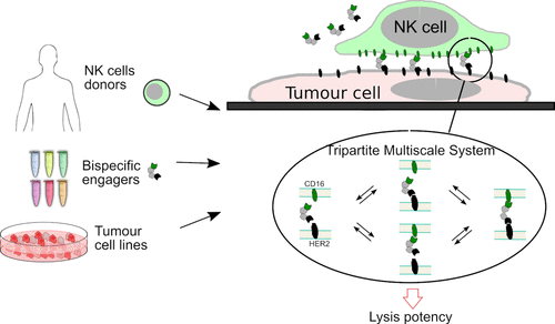
In recent years, immunotherapy has brought about a paradigm shift in the treatment of cancer, making it possible to stimulate and re-arm the immune system against the disease. One strategy already used against certain cancers involves injecting molecular engaging agents (e.g. bispecific monoclonal antibodies) capable of recruiting immune cells such as lymphocytes to directly attack the targeted tumour cells. These bispecific molecules act as a molecular bridge between activating receptors on immune cells and tumour antigens exposed by the target cells. Despite the potential of these new agents, the ability to predict their efficacy remains limited, as it depends on both the patient and the details of the agents molecular structure. Patrick Chames and his team (CRCM) have for several years been developing bispecific antibodies constructed from camelid monodomain antibodies, known as nanobodies.
the authors tested in-vitro a range of 6 original bispecific antibodies combining several nanobodies, and recruiting Natural Killer cells from 15 healthy donors to different target tumour cell lines. They systematically measured the quantity of target cells killed for varying concentrations of engaging molecules, taking into account biophysical parameters such as antibody affinity and receptor density. These data showed that it was possible to largely decouple the role of donor cells from the molecular details of the engager to predict the effective engager dose. They also proposed a simple formula for quantitatively predicting this effective dose in vitro.
Original bispecific engager molecules are being tested to recruit immune cells from healthy donors to kill tumour cells. Donor and molecule parameters are decoupled and the multi-scale model predicts the optimal dose required to kill in vitro (Credit: L. Limozin).
We are pleased to announce the publication of a review authored by Pierre Bongrand in the International Journal of Molecular Sciences. This review examines the growing integration of artificial intelligence into everyday biomedical research and practice, questioning whether this represents a true scientific revolution or a temporary hubris of its potential.
The paper explores:
Insights based on a review of past scientific progress, with an emphasis on immunology, it is concluded that current “omic” data contain useful information that is not adequately interpreted by currently available theoretical processing methods.
A brief description of currently available artificial intelligence tools is presented, with a discussion of expected potential and pitfalls.
Applications of AI in the biomedical field, are described with an emphasis on the biomedical domain.
It is concluded that it is already warranted to apply artificial intelligence to routine biomedical practice, but it is essential to develop validation procedures.


You must be logged in to post a comment.