
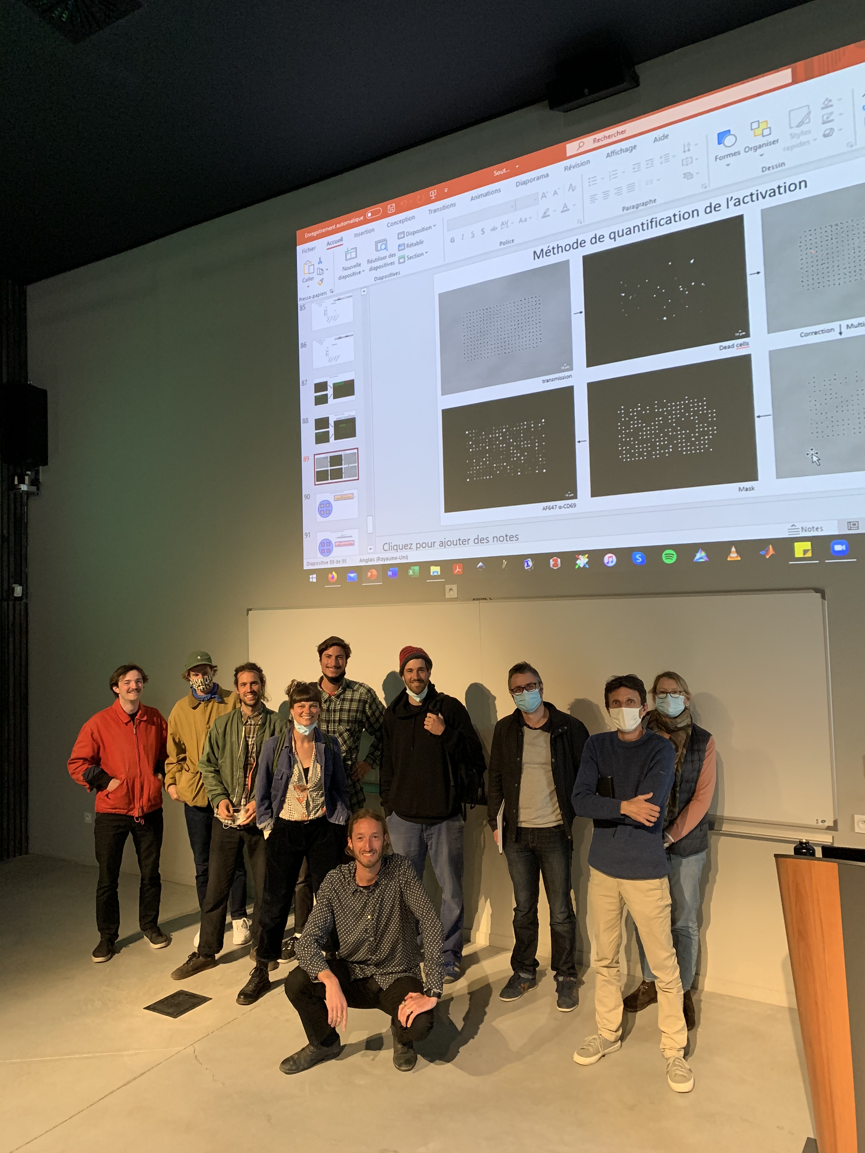
After almost 4 years with us, Farah Mustapha has successfully defended her PhD. Farah completed her work under the supervision of Pierre-Henri Puech at LAI and Kheya Sengupta at CINaM (Centre Interdisciplinaire de Nanoscience de Marseille). Throughout her PhD, she produced soft gels as a substitute for antigen presenting cells (APC) to perform traction force microscopy (TFM) experiments with the aim of investigating the role of mechanotransduction in T cell activation. 🥂
Valentine participated in the AQV DAYS 2022: QUANTITATIVE APPROACHES TO LIVING SYSTEMS conference where she presented a poster entitled “Haptotaxis towards low adhesion” and won the prize for best poster! Congratulations Valentine!
LAI welcomes the new PhD students within its fold. This year, 5 students are joining us.
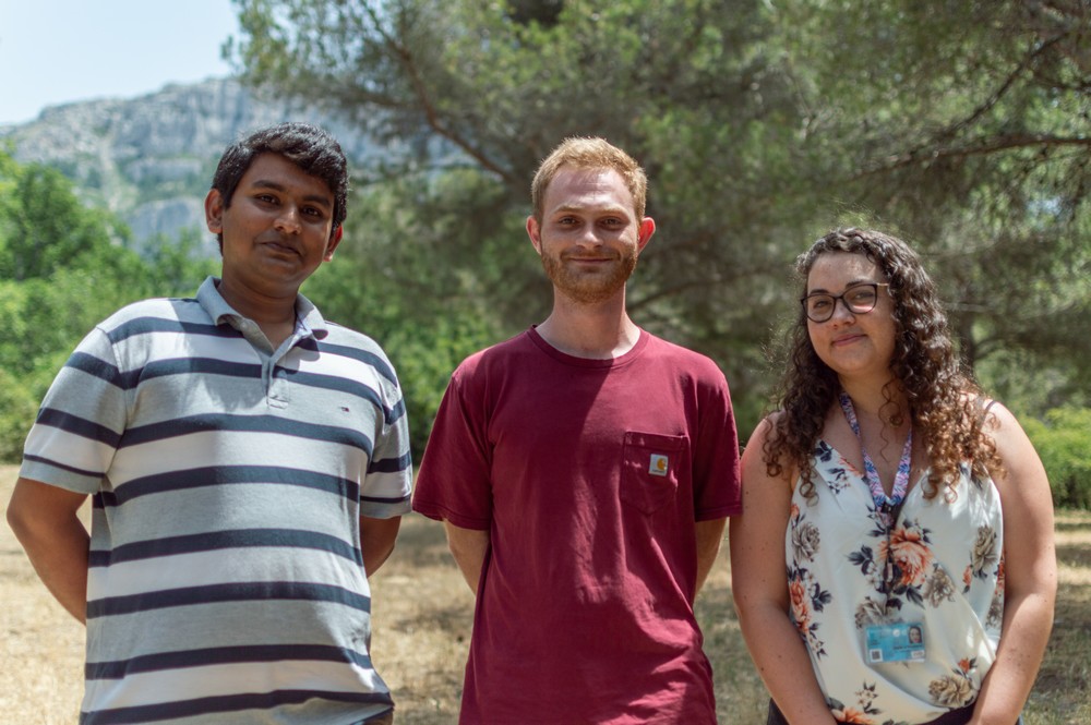
Ismahene Mesbah will be working on decoding protein structural folds, sequence, and structural motifs for the mechanical stability of proteins, and Devam Purohit will be quantifying and manipulating the molecular elasticity of muscle in vitro and in vivo. Both are working at LAI and IBDM and are funded by CENTURI.
We also have Luc David-Broglio who will be developing immunological diagnostic tools that are based on antibody micro-printing and his project is funded by Région PACA, Chandrasekar Subramani-Narayana who will work between LAI and LIS and will use deep learning for image inference, resolution increase and classification of adhered immune cells and is funded by AMU, and finally, Loriane Maillot who will be working between LAI and CIML to investigate molecular dynamics of the thymic immunological synapse during leukemogenesis.
A big welcome to our new batch of students this year, and all the best on your journey!
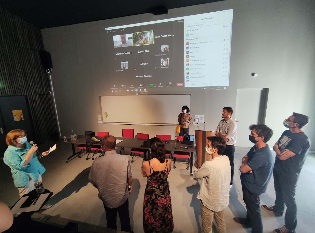
Yet another summer that ends with the graduation of a new PhD. Nicolas Garcia Seyda joined the lab as an engineer before deciding to pursue his PhD in the sunny city. He successfully defended his thesis entitled “Live imaging of leukocyte migration in controlled in vitro setups”. His presence in the lab is surely going to be missed, we wish him the best on his new adventures!
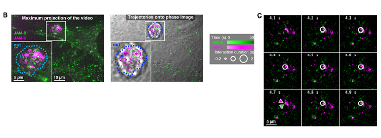
The interactions between haematopoietic and stromal cells are profoundly altered by leukaemias, contributing to the phenomena of resistance to myeloablative treatments. In this study, we followed the dynamics of JAM adhesion molecules at the membrane between leukaemic and stromal cells by videonanoscopy in order to study the establishment and evolution of these cellular junctions. The trajectories of JAMs were analysed with near-nanometer precision using a dedicated MTT (Multi-Target Tracing, Sergé et al. Nature Methods 2008) algorithm extended to 2 colours, which allows to reveal the signature of interaction and stabilization events at cell contacts. We have thus characterised the involvement of JAMs in the interaction mechanisms of tumour cells as well as the maintenance of stem cells in bone marrow niches through enhanced interaction. From a therapeutic perspective, we destabilised leukaemic stem cells using blocking antibodies opening opportunities for disrupting LSC resistance mechanisms.
(B) Maximum projection of a 500-frame videonanoscopy acquisition, showing JAM-B and JAM-C positions over time (left). Maps of JAM-B and JAM-C trajectories represented by gradients of green and magenta, respectively, according to time, and superimposed on the transmission image of the cells (right). Inserts show magnifications of the framed areas. Spatiotemporal colocalizations are denoted by white circles with a size proportional to duration. Several concentric circles correspond to successive colocalizations at a nearby locations but with different durations. (C) Images from the same videonanoscopy acquisition corresponding to the area framed in B, with colocalization events or not (white circle or green/magenta arrowheads, respectively).
This article presents the evidence that immune cells are regulating very rapidely, even before classical signalling times as recorded by calcium fluxes, their mechanical properties when encountering an activating substrate, being either artificial (bead) or physiological (APC). For this, we developed a micropipette-based rheometer to track cell viscous and elastic properties. We have shown that leukocytes become up to 10 times stiffer and more viscous during their activation. Elastic and viscous properties evolve in parallel, preserving a ratio characteristic of the leukocyte subtype. These mechanical measurements set up a complete picture of the mechanics of leukocyte activation and provide a signature of cell function
Activation of three types of leukocytes studied with the micropipette rheometer. Left (a): T cell, middle (b): B cell, right (c): PLB cells. All bars represent 5 μm.
Geoffrey Delhaye has successfully defended his PhD thesis! After 3 years in sunny Marseille, he’s off to a new adventure befitting his free spirit. Our warmest congratulations to you, Geoffrey, and best of luck in your future pursuits!
LAI is happy to welcome the new batch of PhDs joining us this year.
Valentine Seveau will be using micro-fluidic techniques and micro-patterning technologies (PRIMO Alveole) to study the guidance of T cell lymphocytes mediated by integrins and cellular adhesion molecules on a substrate, also known as haptotaxis. She’ll be co-directed by Olivier Théodoly and Marie-Pierre Valignat.
Elodie Lafargue will be trying to characterize for the first time the rest structure of voltage-dependent channels at the molecular level as well as the dynamics of electrical signal transduction when opening or closing the transmembrane channel thanks to two novel breakthroughs in High-Speed Atomic Force Microscopy (HS-AFM) technology. Elodie will be supervised by Ignacio Casuso.
Rémy Torro and Lama Awada also joined us through the CENTURI PhD program. Rémy will be co-directed by Laurent Limozin at LAI and Kheya Sengupta at CINAM, he’ll be using reflection interference contrast microscopy (RICM) and deep learning-based techniques, as a systematic approach to perform 3D reconstructions of cell membranes at the interface with substrates. Lama on the other hand, is supervised by Philippe Robert at LAI and Pierre Milpied at CIML and she’ll be trying to characterize the nature of physical cues driving antibody affinity maturation in B cell immune responses using the laminar flow chamber in 2D while simultaneously integrating parallel analysis of transcriptome and antibody genes sequences at single-cell resolution.
Welcome everyone and we wish you a successful journey!






You must be logged in to post a comment.