A new article on morphodynamics of T-lymphocytes: Scanning to spreading

Binding of the T cell receptor complex to its ligand, the subsequent molecular rearrangement, and the concomitant cell-scale shape changes represent the very first steps of adaptive immune recognition. The first minutes of the interaction of T cells and antigen presenting cells have been extensively scrutinized; yet, gaps remain in our understanding of how the biophysical properties of the environment may impact the sequence of events. In particular, many pioneering experiments were done on immobilized ligands and gave major insights into the process of T cell activation, whereas later experiments have indicated that ligand mobility was of paramount importance, especially to enable the formation of T cell receptor clusters. Systematic experiments to compare and reconcile the two schools are still lacking. Furthermore, recent investigations using compliant substrates have elucidated other intriguing aspects of T cell mechanics. Here we review experiments on interaction of T cells with planar artificial antigen presenting cells to explore the impact of mechanics on adhesion and actin morphodynamics during the spreading process. We enumerate a sequence tracing first contact to final spread state that is consistent with current understanding. Finally, we interpret the presented experimental results in light of a mechanical model that captures all the different morphodynamic states.

https://doi.org/10.1016/j.bpj.2024.02.023

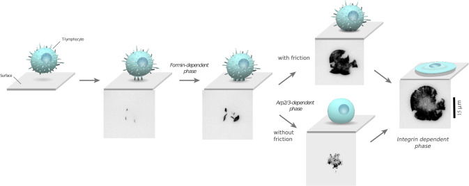
Sequence of ligand recognition and spreading phases. Schemes (not to scale) show T cell morphology, and micrographs show labeled actin cytoskeleton (GFP-lifeact, TIRF mode) as a T cell undergoes adhesion to a ligand-covered surface.

A new article on antigen density and applied force control enrichment of nanobody-expressing yeast cells in microfluidic

In vitro display technologies such as yeast display have been instrumental in developing the selection of new antibodies, antibody fragments or nanobodies that bind to a specific target, with affinity towards the target being the main factor that influences selection outcome. However, the roles of mechanical forces are being increasingly recognized as a crucial factor in the regulation and activation of effector cell function. It would thus be of interest to isolate binders behaving optimally under the influence of mechanical forces. We developed a microfluidic assay allowing the selection of yeast displaying nanobodies through antigen-specific immobilization on a surface under controlled hydrodynamic flow. This approach enabled enrichment of model yeast mixtures using tunable antigen density and applied force. This new force-based selection method opens the possibility of selecting binders by relying on both their affinity and force resistance, with implications for the design of more efficient immunotherapeutics.

https://doi-org.proxy.insermbiblio.inist.fr/10.1039/D4LC00011K

A new article on AI Tools for Immunofluorescence-Based Diagnostics

We are excited to share our latest research published in the International Journal of Molecular Sciences: “Comparison of the Capacity of Several Machine Learning Tools to Assist Immunofluorescence-Based Detection of Anti-Neutrophil Cytoplasmic Antibodies.”

This work was conducted in collaboration with Daniel Bertin and Nathalie Bardin, both members of the Immunology Laboratory of Marseille Hospitals, and Pierre Bongrand from LAI.

This study addresses a current challenge of using artificial intelligence to perform biomedical tasks without any human intervention, which might result in lower cost, increased availability and increased rapidity. Here, we compared the capacity of several widely available machine learning tools to analyze microscopic images in order to diagnose some autoimmunity diseases. The paper described preliminary results obtained with a dataset of 1,733 cell images.

For more information, visit the publication: https://doi.org/10.3390/ijms25063270.

A new article on Naive T lymphocytes chemotax long distance to CCL21 but not to a source of bioactive S1P in Iscience

Naive T lymphocytes traffic through the organism in search for antigen, alternating between blood and secondary lymphoid organs. Lymphocyte homing to lymph nodes relies on CCL21 chemokine sensing by CCR7 receptors, while exit into efferent lymphatics relies on sphingolipid S1P sensing by S1PR1 receptors. While both molecules are claimed chemotactic, a quantitative analysis of naive T lymphocyte migration along defined gradients is missing. Here, we used a reductionist approach to study the real-time single-cell response of naive T lymphocytes to CCL21 and serum rich in bioactive S1P. Using microfluidic and micropatterning ad hoc tools, we show that CCL21 triggers stable polarization and long-range chemotaxis of cells, whereas S1P-rich serum triggers a transient polarization only and no significant displacement, potentially representing a brief transmigration step through exit portals. Our in vitro data thus suggest that naive T lymphocyte chemotax long distances to CCL21 but not toward a source of bioactive S1P.

https://doi.org/10.1016/j.isci.2023.107695

Non-exhaustive model of naive T lymphocyte traffic after accessing lymph nodes. Cells are gently attracted by long-range CCL19 and CCL21 gradients toward the central parenchyma, where they encounter higher and homogeneous concentrations of CCL21 that allow their random walk throughout the T cell zone. Moreover, S1P-rich serum triggers a transient polarization only and no significant displacement, potentially representing a brief transmigration step through exit portals.

A new article on OT_Analysis: a software for rich analysis of force curves when probing living cells with optical tweezers

Optical tweezers are a light-based technique for micromanipulating objects. It allows to move objects such as microbeads and cells, and to record minute forces down to a few pN, which makes it a technique very well adapted to mechanical measurements on living cells (Gennerich, 2017). We are interested in the mechanotransduction properties of lymphocytes. We seek to dissect the effect of forces and cell mechanics on the cellular response, in the context of the immune system. T cell mechanotransduction has been recently demonstrated to be instrumental in the finesse and accuracy of the response of the latter Puech & Bongrand (2021)]. In addition, cells can exert forces when performing their action, e.g. cytotoxic T cells are using forces to kill target infected cells (Basu et al., 2016).
Using optical tweezers and specifically decorated beads as handles, we pull membrane nano- tubes from gently adhered living lymphocytes (Sadoun et al., 2020). Such nanotubes are usually used to probe the tension of adherent cells (Diz-Muñoz et al., 2010). By varying the antibodies that are used to decorate the beads, we select the molecule type we specifically pull on, and we then explore the molecules which are characteristic of the immune synapse, which is one of the key organizational structures that have profound implications in T cell recognition and action (Baldari & Dustin, 2017).
Using this approach, we probe not only the forces of recognition of the given antibody to its target molecule, but also, by using strong extracellular bridges, we probe the cytosolic link of the probed molecule to the cytoskeleton. Such a link has been proposed to be instrumental in the way T cells can apply or feel forces through the molecule. A theoretical model has been built and has been recently reported in a dedicated article (Manca et al., 2023). Furthermore, we will demonstrate the application of the software on full data.

The experimentally obtained data consists of force signal as a function of time (among other parameters), in the three directions of space, obtained in large quantities (at least 10 per cell / bead couple, and up to 20 couples tested per sample), containing rich and detailed features that can relate to molecular and/or cellular mechanics that our model explores. It is therefore needed to standardize and semi-automate data analysis to help the experimentalist, often a biologist, to extract relevant features from the experimental data sets.

https://doi.org/10.21105/joss.04877

A new article on probing mechanical interaction of immune receptors and cytoskeleton by membrane nanotube extraction

The role of force application in immune cell recognition is now well established, the force being transmitted between the actin cytoskeleton to the anchoring ligands through receptors such as integrins. In this chain, the mechanics of the cytoskeleton to receptor link, though clearly crucial, remains poorly understood. To probe this link, we combine mechanical extraction of membrane tubes from T cells using optical tweezers, and fitting of the resulting force curves with a viscoelastic model taking into account the cell and relevant molecules. We solicit this link using four different antibodies against various membrane bound receptors: antiCD3 to target the T Cell Receptor (TCR) complex, antiCD45 for the long sugar CD45, and two clones of antiCD11 targeting open or closed conformation of LFA1 integrins. Upon disruption of the cytoskeleton, the stiffness of the link changes for two of the receptors, exposing the existence of a receptor to cytoskeleton link—namely TCR-complex and open LFA1, and does not change for the other two where a weaker link was expected. Our integrated approach allows us to probe, for the first time, the mechanics of the intracellular receptor–cytoskeleton link in immune cells.

https://doi.org/10.1038/s41598-023-42599-9

A new tool was developed at LAI!

In recent work, Yong Jian Wang, Claire Valotteau, and Laurent Limozin managed to combine DNA scaffolds and acoustic force spectroscopy to characterize individual protein bonds. This work has been in done in collaboration with colleagues at CRCM and IBENS.

To know more about the work, don’t hesitate to reach out to Laurent Limozin or take a look at the article: https://www.cell.com/biophysj/fulltext/S0006-3495(23)00302-8

In press: CNRS

© Vladimir Kunetki – CNRS

A new article on living cells guided against a flow by a roly-poly-like mechanism in PNAS

Cellular guidance by chemical or physical signals is essential for many life processes and usually relies on sophisticated biological processes that are still partially elucidated. Microfluidic experiments and mechanical modeling has revealed that the choice for cells to orient themselves against or in the direction of a flow can result from a simple physical bias. They have worked with  keratocytes, cells that form the scale of fishes, and whose morphology is characterized by broad  flat “front” and a compact protruding front  “back”. A simplified model of a cell with a hemispherical back and a flat rectangular front allows to quantitatively calculate the forces that the flow exerts on each edge. The resulting force stabilizes the cells with a large rear edge against the flow, like a roly-poly that stays upright because of its heavy bottom edge. The researchers’ model successfully predicted the experimentally observed orientation for each cell without adjusting parameter. It is an elegant example where a characterized biological behavior does not result from specialized molecular sensors and a complex cascade of internal biosignals to reorient the cell, but from a simple passive physical bias.

https://www.pnas.org/doi/10.1073/pnas.2210379119

A– Keratocytes descending a flow, the white arrow indicates the direction of the stream. B– Cell morphology seen in 3D by confocal microscopy, with a bulbous back edge and a flat, thin front edge. C– Cell modeling with a hemisphere at the back (red) and a flat rectangle at the front (brown) D– The torques resulting from flow on cell front (red arrow) and cell front (brown arrow) stabilize upstream orientation of cell with larger rear edge, like the torque resulting from gravity stabilizes the standing position of roly-poly toys with large bottom edge. 

This paper was commented on in CNRS (link)

A new article on leukemic stem cell adhesion to bone marrow stromal cells by single-molecule tracking nanoscopy in Journal of Cell Science

The interactions between haematopoietic and stromal cells are profoundly altered by leukaemias, contributing to the phenomena of resistance to myeloablative treatments. In this study, we followed the dynamics of JAM adhesion molecules at the membrane between leukaemic and stromal cells by videonanoscopy in order to study the establishment and evolution of these cellular junctions. The trajectories of JAMs were analysed with near-nanometer precision using a dedicated MTT (Multi-Target Tracing, Sergé et al. Nature Methods 2008) algorithm extended to 2 colours, which allows to reveal the signature of interaction and stabilization events at cell contacts. We have thus characterised the involvement of JAMs in the interaction mechanisms of tumour cells as well as the maintenance of stem cells in bone marrow niches through enhanced interaction. From a therapeutic perspective, we destabilised leukaemic stem cells using blocking antibodies opening opportunities for disrupting LSC resistance mechanisms.

https://doi.org/10.1242/jcs.258736

(B) Maximum projection of a 500-frame videonanoscopy acquisition, showing JAM-B and JAM-C positions over time (left). Maps of JAM-B and JAM-C trajectories represented by gradients of green and magenta, respectively, according to time, and superimposed on the transmission image of the cells (right). Inserts show magnifications of the framed areas. Spatiotemporal colocalizations are denoted by white circles with a size proportional to duration. Several concentric circles correspond to successive colocalizations at a nearby locations but with different durations. (C) Images from the same videonanoscopy acquisition corresponding to the area framed in B, with colocalization events or not (white circle or green/magenta arrowheads, respectively).

New article on early leukocyte activation triggered by viscoelastic changes in Biophysical journal

This article presents the evidence that immune cells are regulating very rapidely, even before classical signalling times as recorded by calcium fluxes, their mechanical properties when encountering an activating substrate, being either artificial (bead) or physiological (APC). For this, we developed a micropipette-based rheometer to track cell viscous and elastic properties. We have shown that leukocytes become up to 10 times stiffer and more viscous during their activation. Elastic and viscous properties evolve in parallel, preserving a ratio characteristic of the leukocyte subtype. These mechanical measurements set up a complete picture of the mechanics of leukocyte activation and provide a signature of cell function

https://doi.org/10.1016/

Activation of three types of leukocytes studied with the micropipette rheometer. Left (a): T cell, middle (b): B cell, right (c): PLB cells. All bars represent 5 μm.